Home
We are often challenged by research questions about small particles in the ocean: How big are they, what are they, and how do they move. These are tricky questions to answer because accurate measurement of small particles suspended in the water is technically demanding due to their complicated, delicate structures which cover a wide range of sizes, concentrations, and movement characteristics.
Since 2014 we have been developing new in-situ particle imaging techniques to help answer questions surrounding the nature of suspended particles in the marine environment. Conventional lens-based imaging of suspended particles often suffers from limitations related to depth-of-field, particle occlusion, and perspective errors. These errors can be overcome by the use of holographic imaging or restricted path length telecentric systems such as the SINTEF SilCam.
An example summary of particle images and statistics that are obtained from PySilCam:

SINTEF's SilCam has been developed in-house within the department of Environmental Technology, initially to measure large particulate pollutants in seawater. The system has been used both in large-scale experimental subsea releases of oil and gas, and in the field to quantify the distribution of suspended material such as zooplankton, large phytoplankton, mineral grains and marine snow. Images are analysed to provide size distribution and concentrations spanning from ~50𝜇𝑚 to several cm in length, and an approach utilising Deep Convolutional Neural Networks (Tensorflow) is used to identify the type of particle, be it a copepod, diatom chain, marine snow etc. True, in-focus particle images are recorded in colour directly, so minimal processing is needed. These images look very much like microscope images (albeit in a lower magnification). A typical image (of 15MB) is processed in about 0.7 seconds to retrieve a particle size distribution, concentration and classified particle types within the image.

The SilCam is under continuing development to address the ever-evolving research questions within marine environmental research.
Past developments have enabled (and continue to support) research on the following topics:
- Quantifying the effect of subsea dispersant injection on oil droplet and gas bubble size distributions.
- Quantifying the effect of high pressure on the efficiency of subsea dispersant injection applied to mixed oil and gas releases.
- Mapping characteristics of particles surrounding a submarine mine tailings discharge. An interactive map of Frænfjorden, with SilCam, LISST and Turbidity profiles can be found here.
- Measurements of gas bubble size distributions from subsea gas releases.
- Automatic identification of salmon lice.
- Automated subsea dispersant injection.
- In-situ observations to support biogeochemical ocean models.
- Enhancing capabilities for subsea blowout monitoring.
Ongoing activity relating directly to development of the SilCam include the following applications:
- Autonomous measurements of meso-zooplankton onboard AUVs.
- Autonomous resource mapping of fish eggs, larvae, and copepods via combined imaging and acoustic monitoring.
- New technology for real-time monitoring and modelling of drilling discharges.
- Improving understanding of the nature and movement of marine oil snow.
- Sediment monitoring relating to deep sea mining
- In-situ settling velocity measurements
- Near-surface particle monitoring
Future challenges we seek to address, include:
- Autonomous resource mapping of fish eggs, larvae, and copepods via combined imaging and acoustic monitoring.
- In-situ salmon lice monitoring and warning systems.
- Microplastic detection and monitoring.
- Behaviour of meso-zooplankton within thin layers.
- In-situ identification of sub-lethal toxic effects on meso-zooplankton.
- Lagrangian monitoring
- Environmental impact monitoring surrounding marine renewable energy devices
The original method for basic analysis of size distributions is described here: Davies, E. J., P. J. Brandvik, F. Leirvik, and R. Nepstad. 2016. The use of wide-band transmittance imaging to size and classify suspended particulate matter in seawater. Mar. Pollut. Bull. doi:10.1016/j.marpolbul.2016.11.063
Elements of this method have been updated since the publication of the original paper. The main methodological difference is that TensorFlow is now used to classify particles.

The following main processing steps are applied to each image recorded by the silhouette system:
- Each image is corrected by a clean background to reduce noise
- The corrected image is segmented (binarized) to produce a logical image (zeros and ones) of the particles detected
- Particles in the binary image are then counted and particle properties (geometry and particle type) are calculated for each particle
- The particle size distribution is calculated by counting Equivalent Circular Diameters (ECD) into their appropriate volume size class (this is done in a similar way to the log-spaced size bins used by Sequoia Scientific's LISST and LISST-HOLO (http://holoproc.marinephysics.org) instruments, but extending up to a maximum diameter of several cm).
The background image used for correction of each raw image is calculated from an average of images recorded immediately prior to the raw image being processed. This allows for real-time moving average background correction. The correction of images reduces noise and gradients in background illumination and small fouling artefacts that may appear of the optics.
The background correction is performed such that corrected pixel values retain sensible physical units, and thus pixel intensities within areas of a corrected image can be (approximately) related to attenuation. The shape of the approximated transmitted spectra (albeit limited to the RGB channels of the camera) can be used as a more quantifiable metric for certain types of material, which can be related to other measurable optical properties and remote sensing.
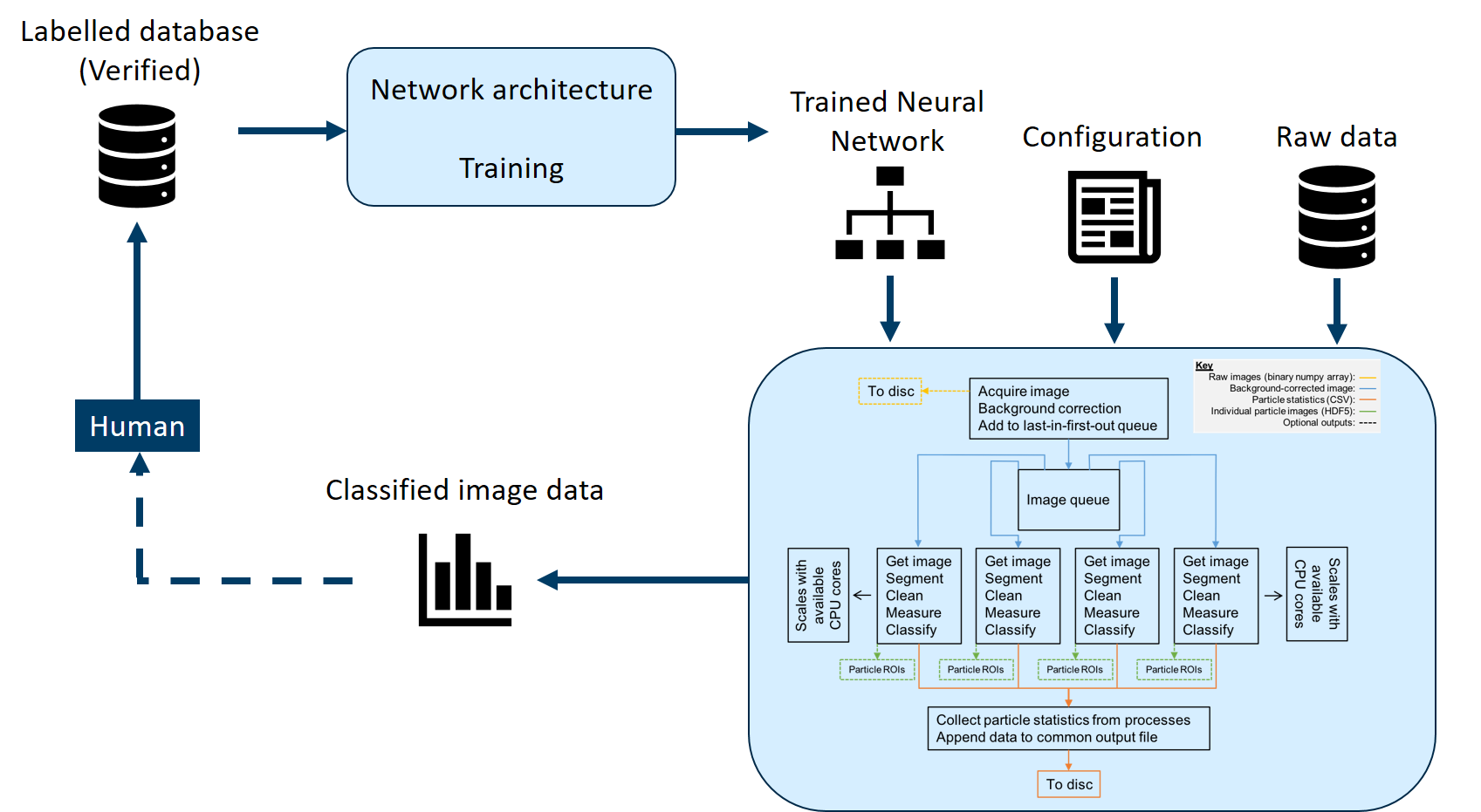
Here is an outline of the workflow used to classify particle types using a the convolutional neural network in PySilCam:
An example of how this type of classification can differentiate eggs, larvae and copepods:

Unsupervised T-SNE clustering of SilCam images of eggs, larvae and copepods indicates that there is potential for unsupervised classification of this type of particle data:
