FloWave.US is a MATLAB program for automated ultrasound blood flow analysis. FloWave.US extracts blood velocity and vessel diameter from ultrasound screen captures, giving researchers the flexibility to calculate a variety of vascular health parameters.
Please visit the wiki for instructions and tutorial links.
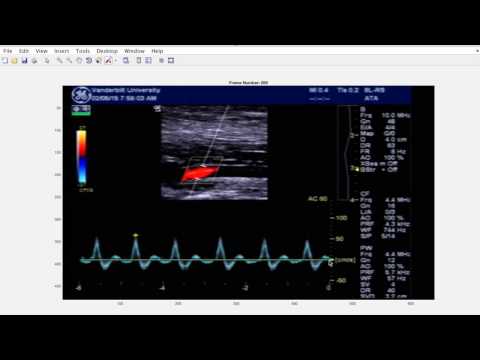
The following video also demonstrates how to analyze the muscle contraction demo video.
- Journal of Applied Physiology Reference
- Example Ultrasound Recordings
- Release Notes
- Getting Started with MATLAB
Assumes MATLAB (release 2011b or newer) installed:
- CLICK the green "Clone or download" button.
- SELECT the "Download ZIP" option to download the latest program release.
- SAVE the unpacked source code files into a folder directory of your choosing.
- OPEN MATLAB and set the working directory to the location of your downloaded files.
- TYPE
FloWaveUSinto the MATLAB command prompt to run the program.
- Assumes an ultrasound screen setup file exists - see the next section for more details.
If you have any installation problems, please file an issue with a description of your problem, and we will try to help you.
FloWave.US requires an ultrasound screen setup file to adapt the program to different ultrasound scanners and operation modes. To create a screen setup file:
- OPEN MATLAB and set the working directory to the location of your downloaded files.
- TYPE
UsSetupinto the MATLAB command prompt and follow the on-screen instructions. - SAVE the ultrasound screen setup file (e.g. "SETUP.mat") in the same working directory for easy access.
FloWave.US analyzes digital video recordings of duplex or brightness mode (B-Mode) ultrasound screen captures. For the best results, the screen capture should meet the following requirements:
- High Definition Acquisiton: video recordings of the ultrasound screen should be acquired at a mininmum sampling rates of 30 frames per second. Video recording equipment can typically be connected to the ultrasound scanner's video output or composite output port.
- Example Recording Equpiment:
- View of On-Screen Scales: screen captures must include the manufacturer's calibration scales (e.g. velocity, distance, time) to convert pixels into the units of interest. These scales should be visible in addition to a complete view of the B-Mode or duplex image.
- MATLAB Compatible Video File: digital video files must be saved/converted to an AVI or MOV format.
- Windows 7 or later: AVI or MOV
- Macintosh: MOV only
- Removal of On-Screen Calculations/Markings: on-screen calculations (e.g. cardiac cycle gates) should be turned off during the video recording to avoid obstructing the view of the B-Mode or duplex image.
- Real-Time Video: video recordings of previously captured data (e.g. cine loops) can cause artifacts or missed frames when reading the video file into FloWave.US. It is possible to analyze these types of data, but it can be a challenge to account for errors. For best results, we recommend recording the ultrasound screen in real-time to a file during an experiment.
We have provided example ultrasound video recordings to ensure the FloWave.US source code works on your computer:
- DemoMuscleContraction.mov: duplex ultrasound video recorded in the lower leg during a 1-s muscle contraction experiment - a test case for the main FloWaveUS.m program or to practice creating ultrasound screen setting files with UsSetup.m.
- DemoBMode.mov: B-Mode ultrasound video recorded in the upper arm at rest - a test case for the vessel diameter measurement program BMode.m.
- GELogiqBookeDefault.mat: ultrasound screen settings file for a General Electric Logiq Book e ultrasound scanner - this file is needed to analyze the muscle contraction demo video.
Have an idea for FloWave.US? Found a bug? Please file an issue on GitHub.
If you create an issue, templates are provided to report bugs and request features. Please be sure to include other context (e.g. ultrasound hardware, video recording equipment) if reporting a problem with the code.
We also encourage community members to contribute to this project. Please read the contributing guidelines to learn more about how you can help.
Please be aware that we support a positive social environment for the FloWave.US community. As such, community members are expected to adhere to the project's code of conduct to facilitate constructive, collaborative behavior.
- FloWave.US is licensed under the MIT license.
- Third-party software (ginputc.m and imgaussian.m) are used according to their respective license agreements.
Thank you for your interest in FloWave.US. We hope the program continues to improve and becomes a community effort to provide high quality, efficient, and inexpensive data processing solutions for ultrasound blood flow research.
If you find FloWave.US useful in your research, please consider citing the following:
- Coolbaugh, Crystal L., Bush, Emily C., Caskey, Charles F., Damon, Bruce M., and Towse, Theodore F., "FloWave. US: validated, open-source, and flexible software for ultrasound blood flow analysis." Journal of Applied Physiology 121.4 (2016): 849-857.