The goal is to build a product that helps doctors quickly identify cases of pneumonia in children.
As such, this project is designed to test your ability to build a labeled dataset that distinguishes between healthy and pneumonia x-ray images; this can be used by ML engineers later on down the line to build a classification product. Your main task will be to create a data labeling job using Appen's platform
This project is designed to build a labeled dataset that distinguishes between healthy and pneumonia x-ray images; this can be used by ML engineers later on down the line to build a classification product.
This dataset is a modified version of Kaggle chest x-ray dataset, with most labels removed. Every piece of data is a chest x-ray image. You may see images taken that are slightly different in size and taken under slightly different exposure times. A typical, labeled image is shown below.
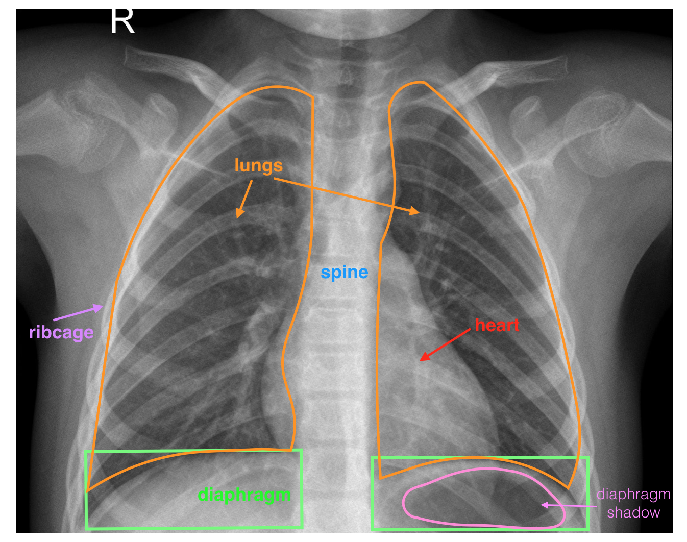
A labeled, healthy, chest x-ray image. Pay close attention to the two lungs and diaphragm (below the lungs)
This is a challenging task because it is not always clear when pneumonia symptoms are present or not in an image. As such, your system is not meant to be a replacement for a doctor, only to aid in quickly identifying healthy patients and surfacing potential cases of pneumonia.
There are a few different visual symptoms that indicate pneumonia. The most important areas to have annotators pay attention to are the lungs and the diaphragm.
- A normal, healthy image will depict clear lungs without any areas of abnormal cloudiness/opacity; there may be structured, web-like vasculature in the lungs but otherwise that area should be clear. In healthy images, you are also more likely to see a diaphragm shadow.
- A pneumonia image may include a few things: areas of cloudiness/opacity in several concentrated areas or one large area. You may also see a general pattern of opacity that obscures the structure of the lungs, heart, and diaphragm.
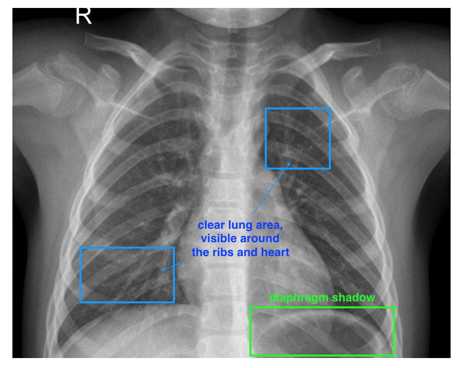
Some characteristics of a healthy image: a clear lung area
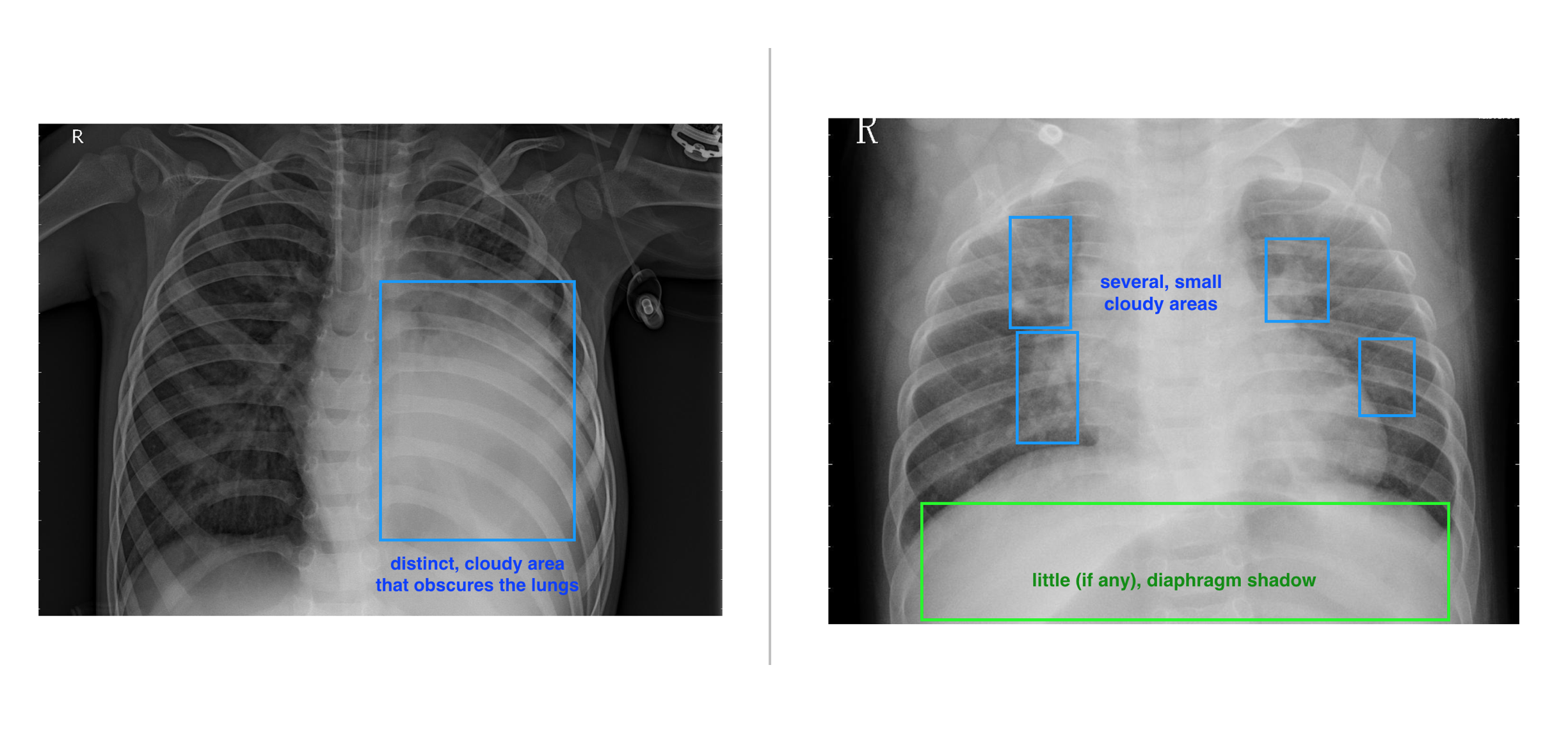
Examples of pneumonia symptoms: (Left) a concentrated, opaque area in the lungs, (Right) multiple, smaller opaque areas throughout the lung area and any diaphragm shadow is obscured
- Can help flag serious cases
- Quickly identify healthy cases
- And, generally, act as a diagnostic aid for doctors