Neurite OASIS Sample Data
Organized data collection including 414 subjects from the open-access OASIS dataset processed with FreeSurfer and SAMSEG for the neurite package.
These data were prepared by Andrew Hoopes and Adrian V. Dalca for the following HyperMorph paper.
If you use this collection please cite the following and refer to the
OASIS Data Use Agreement.
-
Learning the Effect of Registration Hyperparameters with HyperMorph.
Hoopes A, Hoffmann M, D. N. Greve, Fischl B, Guttag J, Dalca AV.
MELBA 2022. -
Open Access Series of Imaging Studies (OASIS): Cross-Sectional MRI Data in Young, Middle Aged, Nondemented, and Demented Older Adults.
Marcus DS, Wang TH, Parker J, Csernansky JG, Morris JC, Buckner RL.
Journal of Cognitive Neuroscience, 19, 1498-1507.
In addition, we provide white matter meshes, which were processed for work in topofit. If you use these meshes, please cite the following:
- TopoFit: Rapid Reconstruction of Topologically-Correct Cortical Surfaces.
A. Hoopes, J. E. Iglesias, B. Fischl, D. Greve‡, A. V. Dalca‡
MIDL: Medical Imaging with Deep Learning. Accepted. 2022.
Download v1.0 here (6.6G) – md5: 081392a8150ff99ab7a64a9ded377835
Download v1.0 here 2D only (24M) – md5: c9ae5864f250c7e4b8d83a104e51ae8e
Download WM meshes for V1.0 (4.5 G) – md5: a2d0f23702ad8094b9074890df7d24df
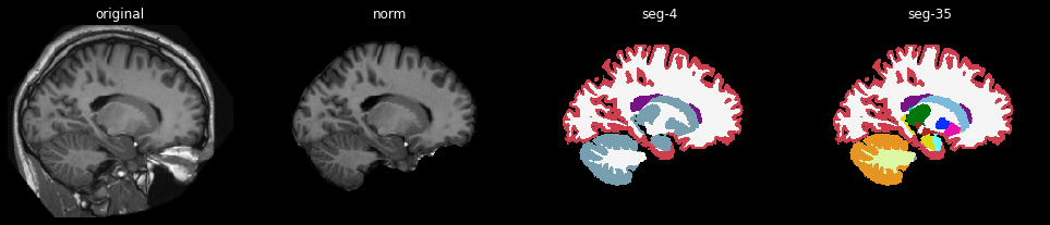
Each subdirectory contains original and normalized T1 scan data as well as
label segmentations for an individual subject. The list of subject IDs can
be found in the subjects.txt file.
Each subject directory has raw (orig) and skull-stripped / bias-corrected (norm)
images in the original scanner space and resampled into an affinely-aligned, common
template space. Additionally, we provide an aligned coronal-slice for 2D applications.
In summary, each subject directory contains the following images:
FILENAME              SHAPE             SPACE

orig.nii.gz           256 x 256 x 256   raw image in scanner space
norm.nii.gz           256 x 256 x 256   corrected image in scanner space

aligned_orig.nii.gz   160 x 192 x 224   raw image aligned in template space
aligned_norm.nii.gz   160 x 192 x 224   corrected image aligned in template space

slice_orig.nii.gz     160 x 192         2D raw image aligned in template space
slice_norm.nii.gz     160 x 192         2D corrected image aligned in template space
We conformed all images to a common shape and scaled them between 0 and 1, and skullstripped and
bias-corrected 'norm' images with freesufer. We
registered and resampled the images into freesurfer's talairach space using the talairach.xfm atlas
transform generated by recon-all and cropped via [(48, 48), (31, 33), (3, 29)]. We also extracted
coronal slice 109 from these cropped volumes to generate the 2D slice images.
Each subject contains a set of corresponding automated label segmentations. We provide a 35-label segmentation of major anatomical regions as well as a 4-label tissue-type segmentation, generated from the former. For the 2D images, we instead provide a 24-label segmentation, consisting of the most common structures found in that sliced region. Corresponding mappings from label ID to structure name are available in the following files:
seg35_labels.txt
seg24_labels.txt
seg4_labels.txt
These are provided in freesurfer's colortable file format, which can be used to visualize the segmentations correctly in freeview. For example:
cd OASIS_OAS1_0445_MR1
freeview norm.nii.gz \
         seg4.nii.gz:colormap=lut:lut=../seg4_labels.txt \
         seg35.nii.gz:colormap=lut:lut=../seg35_labels.txt