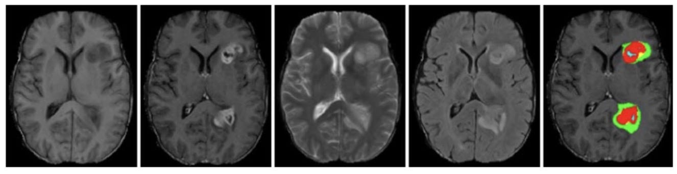
Neural network to automatically segment tumor regions in brain of MRI images
-
Used 484 training images from Decathlon 10 Challenge dataset and
nibabelto extract the images and labels from the files. -
Generated "patches"of our data which are as sub-volumes of the whole MRI images in order to speed up training time and reduce the memory needed
-
Standardized the values to have a mean of zero and standard deviation of 1 to reduce the range of MRI images
-
Built a
3D U-netmodel that takes advantage of the volumetric shape of MR images to predict the regions affected by Edema,Non-enhancing and Enhancing tumor -
Used Dice Similarity Cofficient then Soft Dice as a loss function to face the heavy imbalance in segmentation and
-
Convert prediction from probability into a category by using a
threshold of 0.5 -
Evaluated model's performance for by calculating the sensitivity, specificity