In India and over the world, Cancer has become a deadly disease and more and more people are suffering from Cancer and a survey says one in every 30 women suffer from this disease in their lifetime and so basically the project was first thought of because of the increase in cases of breast cancer and one thing which is very important that if we can detect the Cancer at an early stage then there is an increased chances of it getting cured.So this project lays a foundation in making the detection of the cancer automated so that more and more people can get it diagonised early so as get cured.
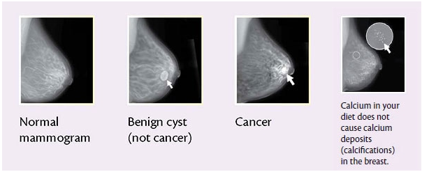
The signs of detection are Masses and micro calcification clusters which are important in early detection of breast cancer.
Micro calcification are nothing but tiny mineral deposits within the breast tissue. They look similar to small white colored spots. They may or may not be caused by cancer.
Masses can be many things, including cysts (fluid-filled sacs) and non-cancerous solid tumors, but they could also be cancerous.
The difficulty in cancer detection is that the abnormalities from normal breast tissues are hard to read because of their subtle appearance and ambiguous margins.Automated tools which can help radiologist in early detection of breast cancer.
Further we have classified the cancer into three categories after its detection- Normal, Malignant, Benign.
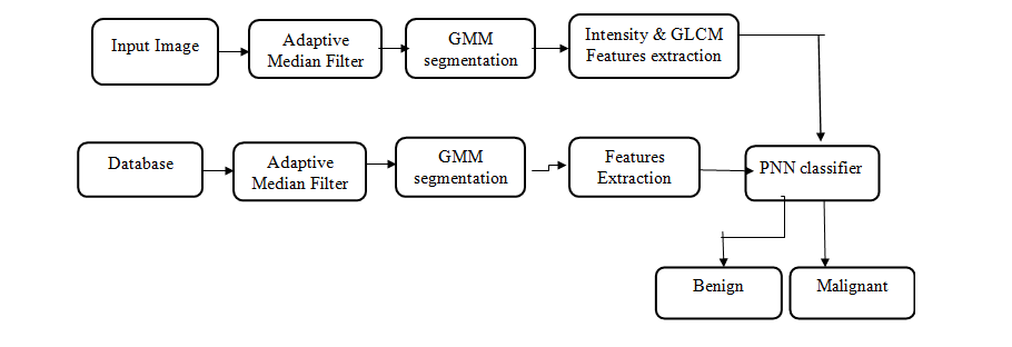
We have used adaptive mean filter to remove noise from image. since it is better among all the spatial filters and distinguish fine details from noise. The Adaptive Median Filter performs spatial processing to determine which pixels in an image have been affected by impulse noise. The Adaptive Median Filter classifies pixels as noise by comparing each pixel in the image to its surrounding neighbor pixels.
The size of the neighborhood is adjustable, as well as the threshold for the comparison. A pixel that is different from a majority of its neighbors, as well as being not structurally aligned with those pixels to which it is similar, is labeled as impulse noise.
These noise pixels are then replaced by the median pixel value of the pixels in the neighborhood that have passed the noise labeling test.we are initially converting the image into grayscale image using rgb2gray() function then applying adaptive mean filtering to the resulting image and then converted the image into unsigned integer 8 using unit8() function.
In this way we preprocessed image.then we performed GMM segmentation(Gaussian Mixture Model) on the preprocessed image with number of regions 2 and number of GMM components 2 and maximum number iterations 10. we performed k-means segmentation with k=2. then we Implemented HMRF-EM (Hidden Markov Random Field Model) and its Expectation-Maximization Algorithm.
The picture decribes the difference between Malignant and Benign tissues in Breast
Open the project in matlab and then run guidemo.m and then a gui mode window will open and then just follow the steps there.For further information check the screenshots.
NOTE--> To get this project working, kindly install MATLAB's Wavelet Toolbox
-
STEP 1: Now you have to browse the image of the mammograms and give it as an input

-
STEP 2: In this step adaptive mean filtering is done

-
STEP 3: GMM Segmentation is done

-
STEP 4: So you can see one as the output in the right side which depicts that the cancer is benign


If you use this work in your research, please cite it as follows:
Sharma, V., Rajasekaran, R. K. & Badhrinarayanan, S. (2019). Visualization of Data Mining Techniques for the Prediction of Breast Cancer with High Accuracy Rates. Journal of Computer Science, 15(1), 118-130. https://doi.org/10.3844/jcssp.2019.118.130
Copyright: © 2019 Vasudev Sharma, Raj Kumar Rajasekaran and Shreya Badhrinarayanan.