Data
Each file is a 512*512 TIFF image, separated into folders, where each folder is a single sample. There are 19 training samples, and 9 testing samples.
Each folder contains a variable number of images. For example, sample 00.00 contains 3,024 images, while sample 00.01 contains 3,048. The image files themselves are numbered, e.g. image00000.tiff, but all the images in a single folder represent the same sample, just taken at different times with different calcium levels. The training labels exist at the sample level, so you’ll use all the images in a single folder to learn the locations of the neurons. Each folder will have a unique sample with unique numbers and positions of neurons. However, while time is a dimension to the data, you may not need to explicitly model it; you’re just interested in finding the active neurons in space.
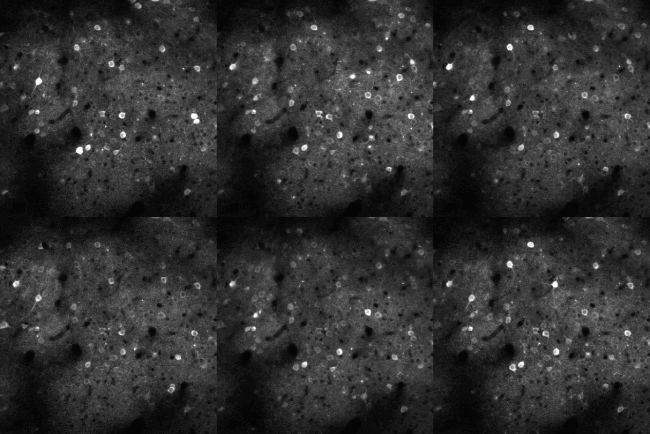
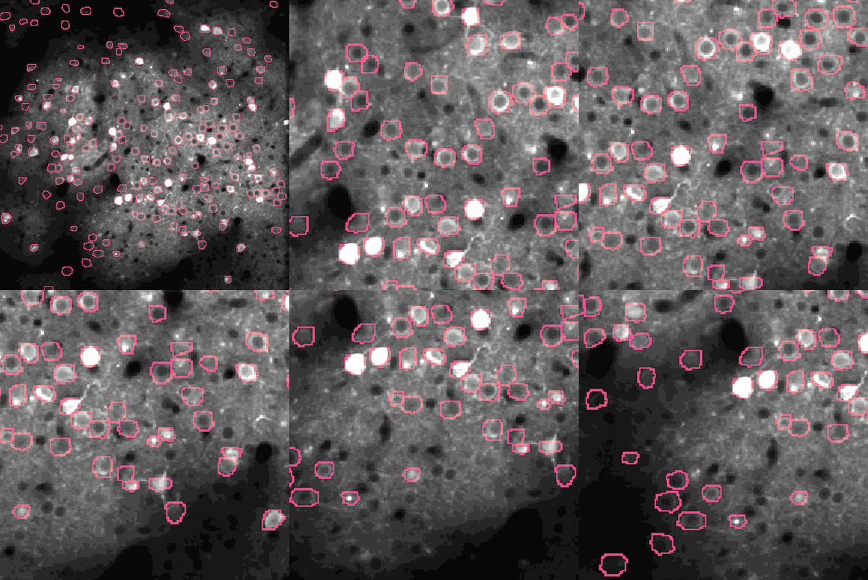
The image on the top represents the time-varying images in the training and testing data. The image on the bottom is the extract "regions of interest" (the pink regions on the right) that correspond to individual neurons, which is the goal to draw circles around the regions that contain neurons.